Footprints Science: Breathing
See breathing in action using this animation, then answer some questions to make sure you understand the process!
The respiratory system brings oxygen into the body and removes carbon dioxide. Understanding how it works helps us see how the body breathes and keeps cells healthy. Use this resource to learn about the organs and functions of the respiratory system.
We need oxygen to survive. It is an important element used by the body during a process called cellular respiration. Cellular respiration is a chemical reaction where glucose is converted to energy in the form of adenosine triphosphate (ATP). This occurs in the mitochondria, as you have learned when looking at organelles.
ATP is used by cells to carry out their functions. Everything from muscle contraction and nerve impulse transmission, to keeping body temperature steady and building new tissues relies on ATP.
Cellular respiration is given by the following word and chemical equations:
\[\textrm{glucose}+\textrm{oxygen}\rightarrow\textrm{carbon dioxide}+\textrm{water}+\textrm{energy}\]\[\ce{C6H12O6}+\ce{6O2}\rightarrow\ce{6CO2}+\ce{6H2O}+\textrm{ATP}\]
But how do we get oxygen to the cells? First, we have to get oxygen into the bloodstream. This is where the respiratory system comes in.
We need a respiratory system to breathe and maintain healthy oxygen levels in the body. This system of organs and tissues is contained inside the ribcage.
The respiratory tract is the pathway that inhaled (and exhaled) air follows to and from the lungs.
A diagram of the respiratory tract, with each component labelled.
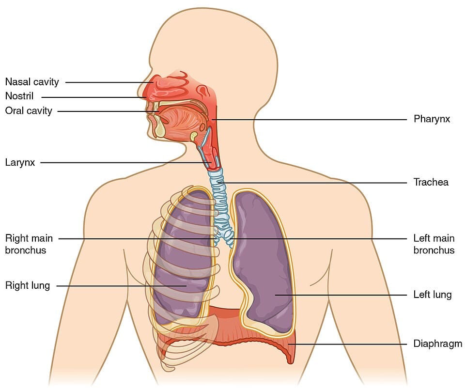
The respiratory tract
Our respiratory tract starts with our nostrils, nasal and oral cavities, and ends with the alveoli deep in our lungs. It works with other organs of the respiratory system, like the diaphragm, to exchange oxygen and carbon dioxide.
Let's look at each part of the respiratory system in more detail.
During the day, your nostrils take turns being more or less congested, and therefore, one lets in more air than the other. You switch "dominant" nostrils every few hours. This is called the nasal cycle.
The oral cavity acts as another route for air intake. It becomes especially helpful during exercise when we need more oxygen.
Together, these cavities make sure that the air reaching our lungs is clean and conditioned for effective gas exchange.
The pharynx, or throat, is a pathway shared by air and food. It connects the nasal and oral cavities to the larynx and oesophagus. In the respiratory system, it guides air down the correct path during breathing.
The larynx sits below the pharynx. You might also know it as the voice box. It brings the air into the next part of the respiratory tract, the trachea, and is also home to the vocal cords which let us speak. The epiglottis, a flap at the top of the larynx, plays a protective role by covering the laryngeal opening during swallowing; this keeps food from entering the airways.
The trachea, or windpipe, is a sturdy tube that goes from the larynx down to the chest. It provides a path for air. The walls of the trachea are surrounded by rings of cartilage (a strong connective tissue), which keep it open and stop the trachea from collapsing during breathing.
At the base of the trachea, the respiratory tract splits into two bronchi. A single one of these paths is called a bronchus. Each bronchus leads to a lung.
A diagram of the trachea and bronchi, with key components labelled.
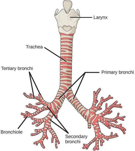
The trachea and bronchi
The cartilage rings around the trachea are not complete; they are like cuffs over the front and sides of the trachea, with an open back. This lets the oesophagus, which sits behind the trachea, expand when you swallow food.
At the end of the bronchioles are clusters of tiny, balloon-like structures called alveoli. Their large surface area, thin walls and proximity to blood vessels allows them to exchange gases within the bloodstream efficiently.
A diagram of an alveoli at the end of a bronchiole, with key components labelled.
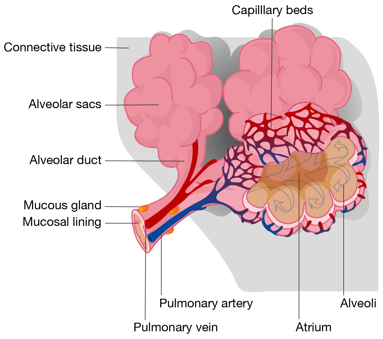
Alveoli
The air we inhale (breathe in) contains a mixture of different gases:
...but we really just need oxygen. So how do we extract just the oxygen we need and get rid of any waste gases? The answer is through a process called gas exchange. This occurs deep within the alveoli of the lungs.
Oxygen from the air we inhale passes through the thin walls of the alveoli into nearby capillaries. The molecules then bind to a protein called haemoglobin, present in the red blood cells, so that they can be transported throughout the body.
Carbon dioxide, a waste product of cellular respiration, passes from the blood into the alveoli to be exhaled (breathed out).
This efficient exchange makes sure that our cells receive the oxygen they need for energy production, while also expelling carbon dioxide to maintain healthy function.
Although nitrogen makes up majority of the air we breathe, it does not play a big role in gas exchange. In fact, nitrogen is an inert gas, meaning that it's not reactive. It is, however, important for us to breathe it in and out anyway, because it helps to keep our alveoli inflated and maintain the pressure balance in our lungs.
The air we exhale is made up of:
We need the help of our muscles to draw breaths in and push them out of our lungs. Most important are the diaphragm and intercostal muscles.
The diaphragm is a large, dome-shaped muscle found at the base of the ribcage. The intercostal muscles are small muscles found between the ribs. The following table summarises how they behave when you inhale and exhale.
| Inhalation | Exhalation | |
|---|---|---|
| Diaphragm | Contracts and moves downward to make more space in your chest cavity so your lungs can expand and fill with air | Relaxes and returns to a dome shape, pushing air out of your lungs |
| Intercostal muscles | Contract to raise the ribcage and expand the chest cavity | Relax and let the ribcage move inward, helping push air out |
One way to remember when the muscles contract and when they relax is to think of inhalation as an active process. Active movement requires muscle contraction. So, these muscles contract when we inhale and relax when we exhale.
Watch this video to see how the organs and tissues of the respiratory system work together to take in oxygen and expel carbon dioxide.
When you breathe, you transport oxygen to the body's cells to keep them working and clear your system of the carbon dioxide that this work generates.
Breathing, in other words, keeps the body alive. So, how do we accomplish this crucial and complex task without even thinking about it? The answer lies in our body's respiratory system.
Like any machinery, it consists of specialised components, and requires a trigger to start functioning. Here, the components are the structures and tissues making up the lungs, as well as the various other respiratory organs connected to them. And to get this machine moving, we need the autonomic nervous system, our brain's unconscious control centre, for the vital functions.
As the body prepares to take in oxygen-rich air, this system sends a signal to the muscles around your lungs, flattening the diaphragm and contracting the muscles between your ribs to create more space for the lungs to expand. Air then whooshes into your nose and mouth, through your trachea, and into the bronchi that split at the trachea's base, with one entering each lung.
Like tree branches, these small tubes divide into thousands of tinier passages called bronchioles. It's tempting to think of the lungs as huge balloons, but instead of being hollow, they're actually spongy inside, with the bronchioles running throughout the parenchyma tissue.
At the end of each bronchiole is a little air sac called an alveolus, wrapped in capillaries full of red blood cells containing special proteins called haemoglobin.
The air you've breathed in fills these sacs, causing the lungs to inflate. Here is where the vital exchange occurs.
At this point, the capillaries are packed with carbon dioxide, and the air sacs are full of oxygen. But due to the basic process of diffusion, the molecules of each gas want to move to a place where there's a lower concentration of their kind.
So as oxygen crosses over to the capillaries, the haemoglobin grabs it up, while the carbon dioxide is unloaded into the lungs. The oxygen-rich haemoglobin is then transported throughout the body via the bloodstream.
But what do our lungs do with all that carbon dioxide? Exhale it, of course.
The autonomic nervous system kicks in again, causing the diaphragm to ball up, and the intercostal muscles to relax, making the chest cavity smaller and forcing the lungs to compress. The carbon dioxide-rich air is expelled, and the cycle begins again.
So that's how these spongy organs keep our bodies efficiently supplied with air. Lungs inhale and exhale between 15 and 25 times a minute, which amounts to an incredible 10,000 litres of air each day. That's a lot of work, but don't sweat it. Your lungs and your autonomic nervous system have got it covered.
The components of the respiratory system and their functions are outlined in the table.
| Component | Function |
|---|---|
| Nasal and oral cavities | Filter, warm and humidify inhaled air |
| Pharynx | Acts as a pathway for air from the nose and mouth to the larynx |
| Larynx | Directs air into the trachea and helps produce sound for speech |
| Trachea | Provides a clear pathway for air to enter and exit the lungs |
| Bronchi | Carries air from the trachea into each lung |
| Bronchioles | Distribute air throughout the lungs to the alveoli |
| Alveoli | Facilitate gas exchange by allowing oxygen and carbon dioxide to move between the air and bloodstream |
| Diaphragm | Contracts and relaxes to change the volume of the chest cavity to facilite inhalation and exhalation |
| Intercostal muscles | Help expand and contract the ribcage to support breathing |
See how well you understand respiration and the components of the respiratory system with a quick quiz.
Read the scenario and use the information provided to answer the questions in the quiz.
Researchers from the Royal Darwin Hospital in the Northern Territory of Australia and the University of Sousse in Tunisia investigated sex differences in pulmonary function among Indigenous Australian with and without chronic airway disease.
The forced vital capacity (FVC) was measured for each individual. This is the maximum amount of air that a person can force out of their lungs after taking in a deep breath.
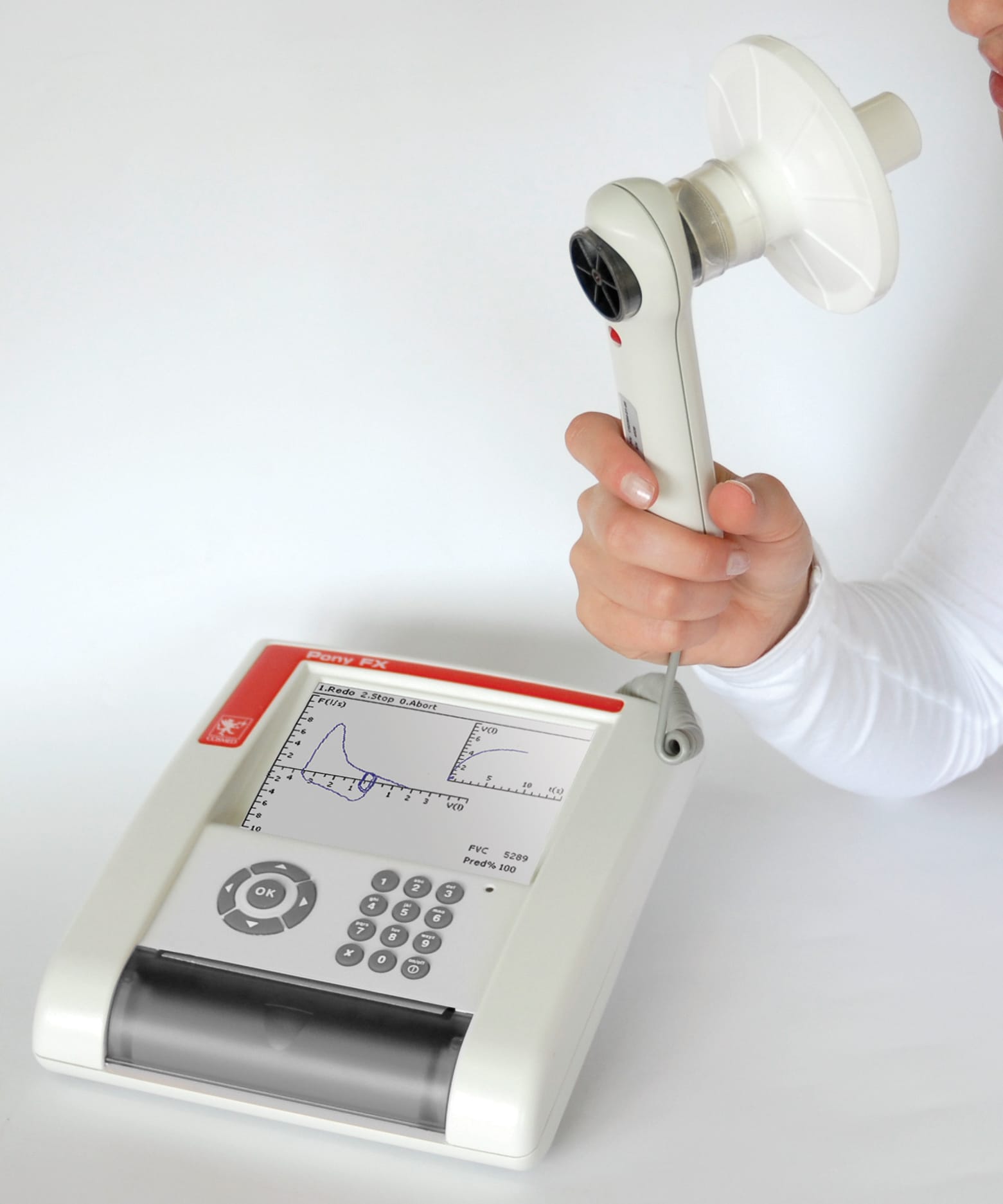
The FVC data for males and females, without chronic airway disease only, is summarised in the table. The p-values are from two-tailed Students t-tests, with p<0.05 considered as statistically significant. The values are averages. The publication also shows the 95% confidence interval for measurements, but they are not shown on this page.
| Female (n=179) | Male (n=111) | p-value | |
|---|---|---|---|
| FVC | 2.59 | 3.63 | <0.001 |
Data from Heraganahally SS, Howarth T, Sorger L and Saad HB (2022), 'Sex differences in pulmonary function parameters among Indigenous Australians with and without chronic airway disease', PLoS One, 17(2):e0263744, doi: 10.1371/journal.pone.0263744, licensed under CC BY 4.0
Images on this page by RMIT, licensed under CC BY-NC 4.0


RMIT University acknowledges the people of the Woi wurrung and Boon wurrung language groups of the eastern Kulin Nation on whose unceded lands we conduct the business of the University. RMIT University respectfully acknowledges their Ancestors and Elders, past and present. RMIT also acknowledges the Traditional Custodians and their Ancestors of the lands and waters across Australia where we conduct our business - Artwork 'Sentient' by Hollie Johnson, Gunaikurnai and Monero Ngarigo.
More information


.jpg)