Action potential
Interested in how the membrane potential changes along the axon during a nerve impulse? Watch this video!
The peripheral nervous system helps the central nervous system communicate with the rest of the body. Learning how it works helps us understand how nerves carry signals to muscles and organs, enabling movement and sensation. Use this resource to learn how the peripheral nervous system keeps us aware and responsive to our environment.
The peripheral nervous system (PNS) connects the central nervous system to the rest of the body, allowing for communication and response. It is the network of sensory organs and neurones that extend from the CNS, reaching every part of the body.
By transmitting signals between the CNS and other regions, the PNS allows our body to respond to stimuli, maintain balance, and coordinate actions, ensuring effective interaction with our environment.
The sensory organs are vital components that allow the nervous system to gather information about the environment. These include eyes, ears, nose, tongue, and skin. Each organ is specialised for detecting specific stimuli.
These organs send signals to the brain, where the information is processed to form perceptions and guide responses. The sensory organs are essential for interacting with and responding to the world.
Human hearing is so sensitive that some people can hear a watch ticking from over \(6\) metres away in a quiet environment!
The longest neurone in the human body connects the base of the spine to the toes. It can be over a metre long!
There are a few types of neurones, but these cells have the same general structure. Their components are summarised in the table.
| Component | Description |
|---|---|
| Cell body (or soma) | Contains the nucleus and other organelles; processes incoming signals and generates outgoing signals |
| Dendrites | Branch out from the cell body; receive signals from other neurones and sensory receptors and pass information along to the cell body |
| Axon | A long, thin projection that transmits signals away from the cell body to other neurones, muscles or glands |
| Axon hillock | Connects the cell body to the axon |
| Myelin sheath | A fatty layer of insulation covering some neurones; helps increase speed of transmission |
| Axon terminals | The end of an axon from which chemicals called neurotransmitters are released |
Neuron by Versal on Sketchfab, licensed under CC BY 4.0
The PNS also consists of ganglia (singular: ganglion), which are clusters of neurone cell bodies. Neurones, on the other hand, are individual signalling cells.
The three types of neurones are:
The sensory and motor neurones are found in the PNS, but the interneurons are found in the CNS, so the brain and spinal cord.
The position of the cell body in these neurones differs, giving them different appearances.
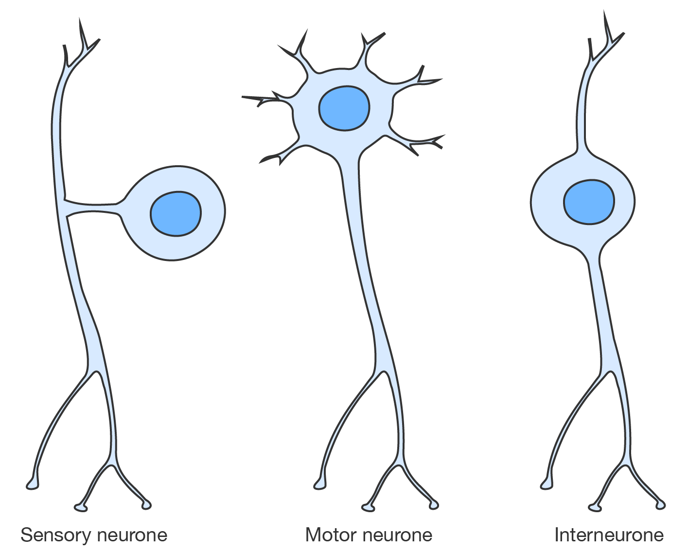
Diagram of a sensory neurone, motor neurone and interneurone
Neurotransmitters are chemical substances that neurones use to communicate with sensory organs, muscles, glands, and other neurones. The location of this communication is called the synapse.
The synapse includes the neurone releasing the neurotransmitters, called the pre-synaptic neurone; the cell receiving the signals, called the post-synaptic cell; and the space between them, known as the synaptic cleft. Neurotransmitters are packaged into little sacs in the pre-synaptic neurone called vesicles.
When an electrical impulse reaches the axon terminal, the vesicles release the neurotransmitters into the synaptic cleft. The neurotransmitters then travel across the gap and bind to specific targets on the post-synaptic cell, called receptors, to pass along the signal.
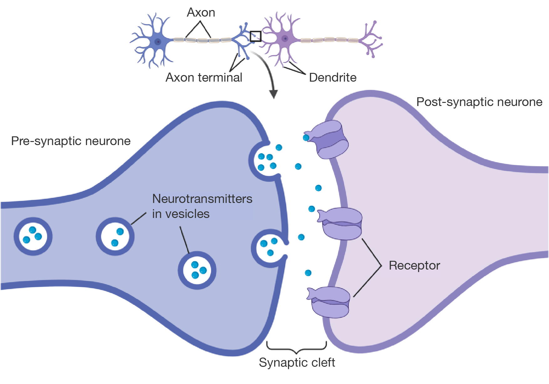
The same neurotransmitter can produce different effects by binding to:
Nerve impulses are electrical signals that travels along neurones. They allow the nervous system to communicate quickly.
The process of generating a nerve impulse can be complicated, so let's break it down.
Watch this video to see how a nerve impulse is propagated through a neurone.
A typical neurone consists of a cell body, plasma membrane extensions called dendrites, an elongated fibre known as an axon, and an axon hillock, the trigger zone that releases a nerve impulse. The axon hillock maintains an excitation limit, or threshold, which determines whether or not a neuron will generate a nerve impulse. A nerve impulse is an electrical signal conducted by a neurone, causing a response in another neurone or target cell. When a neurone is at rest, its membrane is polarised because there are more positive ions outside the cell and more negative ions inside the cell, which creates a charge difference across the membrane. Active transport mechanisms called sodium-potassium pumps carry more sodium and less potassium ions across the membrane to maintain this charged difference. Even in a resting neurone, there is the potential for the charged difference to create an electrical current. This is called a resting membrane potential. When an electrical current flows through a dendrite, this is called a local membrane potential. When a dendrite detects a stimulus, a sodium channel in its plasma membrane opens and lets sodium into the neurone. This influx of positive ions reverses the charge across a particular section of the membrane in a process called depolarisation. To repolarise the membrane, potassium channels open and release potassium out of the neurone. Nearby, a sodium-potassium pump transports excess sodium amount and brings potassium in, which restores the resting membrane potential. The flow of reversing charges along the dendrite's membrane produces a wavelike electrical current toward the neurone's trigger zone. If the strength of the current meets or exceeds the threshold at the trigger zone, an electrical signal called an action potential or nerve impulse will occur. In a nerve impulse, the trigger zone sends an electrical signal down the axon toward the space between neurones called a synapse or to a target cell membrane.
Nerve impulses begin at a sensory organ, which activates a sensory neurone. This stimulates an interneurone, which in turn stimulates a motor neurone. Finally, the motor neurone connects to a target tissue or effector, coordinating it to respond in the required way.
A diagram showing the pathway through which nerve impulses travel.
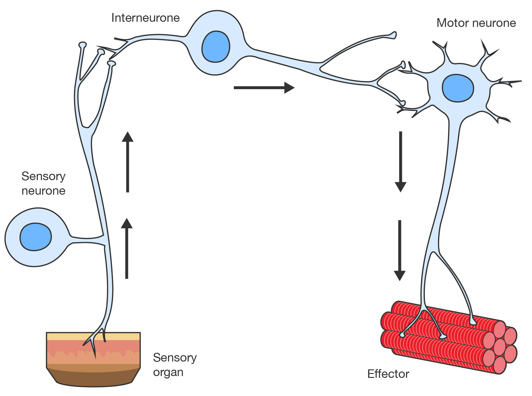
Neurone pathway
The PNS is divided into the somatic and autonomic nervous systems. This division allows the body to efficiently control different types of functions.
The somatic nervous system does this by transmitting sensory and motor signals to and from the central nervous system, allowing us to consciously interact with our environment, move and react.
It is also involved in involuntary movements that are controlled by a reflex arc. One example of a reflex response is pulling your hand away from a hot candle flame.
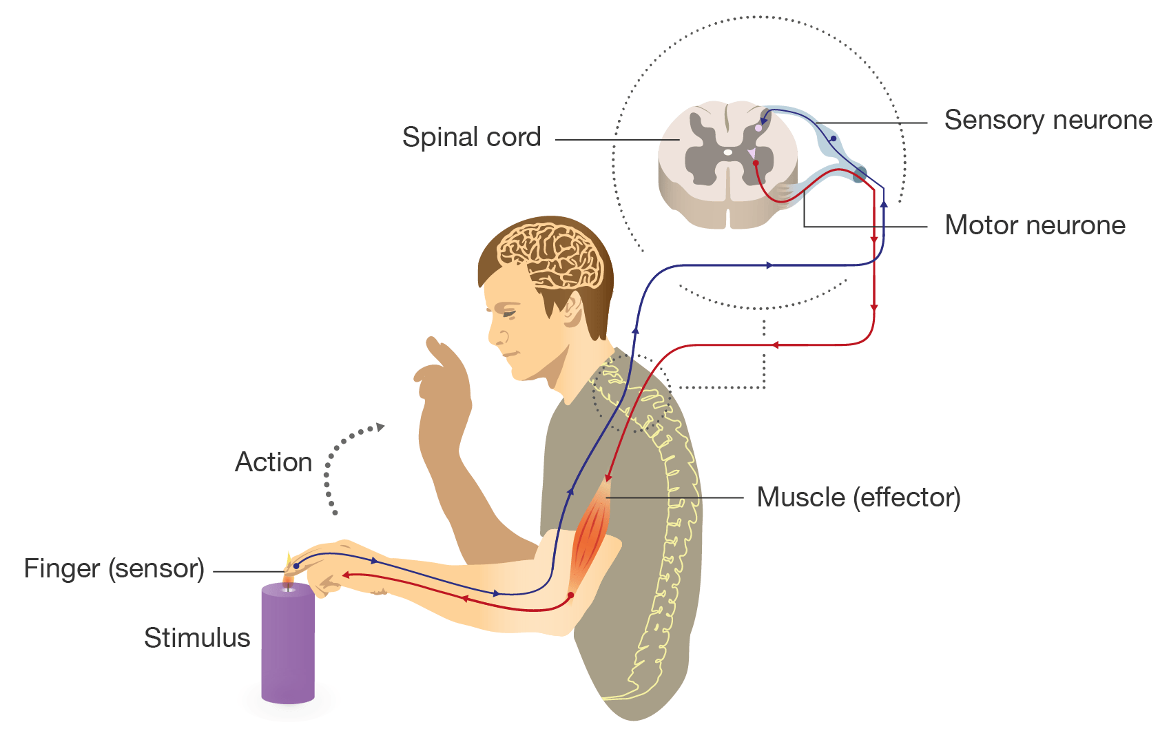
Diagram showing a reflex arc
By operating without conscious effort, the autonomic nervous system ensures vital processes continue smoothly, responding automatically to internal signals and maintaining a state of balance called homeostasis.
The autonomic nervous system can be further divided into the sympathetic and parasympathetic nervous systems.
| Branch | Role | Physiological responses |
|---|---|---|
| Sympathetic nervous system | "Fight-or-flight" during stressful situations |
|
| Parasympathetic nervous system | "Rest and digest" to help body recover |
|
Watch this video to learn more about the sympathetic and parasympathetic nervous systems.
Say you're at the beach on a beautiful day, the Sun is shining, there's a gentle breeze. You dive into the water, but struggle to stay afloat. As you fight the current, your sympathetic nervous system or your fight or flight response kicks in. You are in survival mode and your body prepares for conflict. Your body signals danger by releasing adrenaline. Your heart starts racing. Your pupils expand and your mouth becomes dry. Your airways open up, sending as much oxygen as possible to your brain to keep you alert, and other bodily processes switch gears to help in your time of need. Your stomach and intestines stop digesting, your bladder relaxes, your reproductive system limits blood flow, and your liver releases glucose to give you a burst of energy. All these changes happen so quickly, you may not even be aware of them. When you make it back to shore, you start to calm down and relax. This is your parasympathetic nervous system or your rest and digest process at work. Those bodily processes that were at full attention before change course. Your heart rate slows down, your breathing returns to normal, your pupils constrict and you start to salivate. As your body realises it's no longer under threat, your stomach and intestines begin to digest. Your bladder constricts, and your reproductive system increases its blood flow. In other words, your body returns to its natural state. This is an example of your autonomic nervous system at work. The sympathetic nervous system and parasympathetic nervous system perform a vital balancing act that helps you survive and recover. It happens many times throughout the day, whether you're at the beach, hanging out at home, working from an office or anywhere in between. And while you may tend to think of your fight or flight response kicking in during major stressful situations like a car accident, it can also be triggered by minor things like spilling a glass of milk or getting a paper cut. You can also thank your rest and digest response for all those loving feelings, big and small, you may have from day to day. From cuddling your puppy, to professing your love to your partner. So the next time you are stressed and sense your body tensing up, or you find yourself relaxing on the couch after a busy day of work.
The differences between your sympathetic and parasympathetic nervous systems are so dramatic it can feel a bit like flipping a switch.
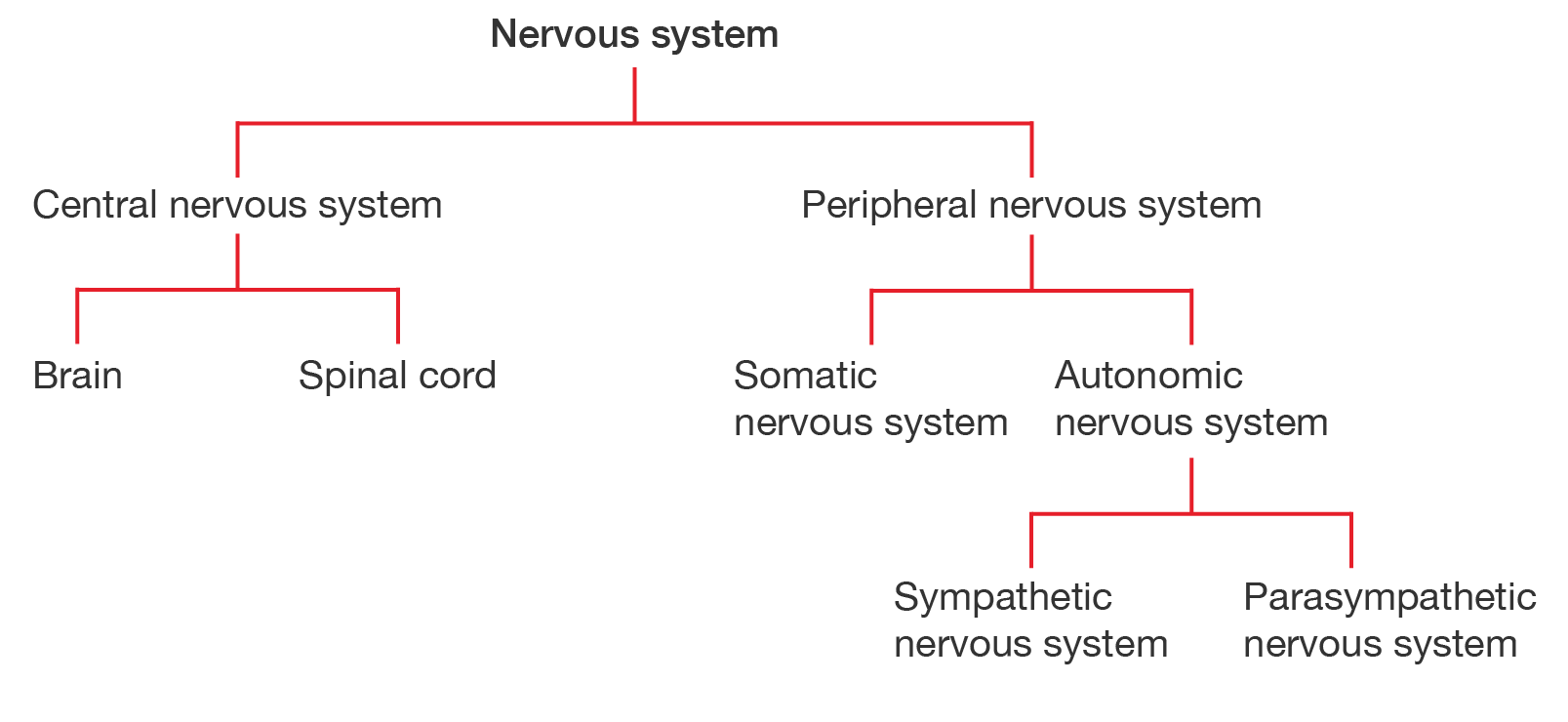
The organisation of the nervous system is summarised in the following flowchart.
See how well you understand the peripheral nervous system with a quick quiz.
Read the scenario and use the information provided to answer the questions in the quiz.
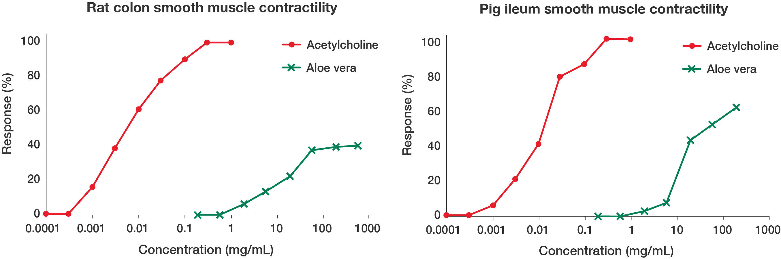
Natural medicines are commonly used to treat constipation. To investigate the potential for processed Aloe vera powder to be used as a treatment, a group of pharmacology researchers from India and Saudi Arabia conducted a study where they treated isolated sections of rat colon and pig ileum with the dissolved powder and measured smooth muscle contraction. They compared the responses to a standard neurotransmitter, acetylcholine, which produces strong contractions and acts as a positive control.
Some of their results are shown in the graphs.
A line graph titled "Rat colon smooth muscle contractility". There are two lines on the graph, a line connecting dots labelled "Acetylcholine" and a line connecting crosses labelled "Aloe vera". The x-axis is labelled "Concentration (mg/mL)", starting at 0.0001 mg/mL and going all the way to 1000 mg/mL. It is represented on a logarithmic scale. The y-axis is labelled "Response (%)", starting at 0 and going to 100, with 20% increments. In the graph, the "Acetylcholine" line starts at 0% reponse for a concentration of 0.0001 mg/mL until a concentration of around 0.0003 mg/mL. Then, the response gradually increases until a concentration of 0.3 mg/mL, where a response of 100% is achieved. From 0.3 mg/mL to 1 mg/mL, the response is 100%. In the graph, the "Aloe vera" line starts at 0% response for a concentration of 0.2 mg/mL until around 0.6 mg/mL. Then, the response gradually increases until a concentration of 60 mg/mL, where a response of around 37% is achieved. From 60 mg/mL to 600 mg/mL, the response seems to mostly plateau, with a maximum of 40% at 600 mg/mL. The data is shown in the table. A similar line graph titled "Pig ileum smooth muscle contractility". The format is the same as the previous graph, but with different data. In the graph, the "Acetylcholine" line starts at 0% reponse for a concentration of 0.0001 mg/mL until a concentration of around 0.0003 mg/mL. Then, the response gradually increases until a concentration of 0.3 mg/mL, where a response of 100% is achieved. From 0.3 mg/mL to 1 mg/mL, the response is 100%. In the graph, the "Aloe vera" line starts at 0% response for a concentration of 0.2 mg/mL until around 0.6 mg/mL. Then, the response gradually increases until a concentration of 200 mg/mL, where a response of around 61% is achieved. The data is shown in the table.
Line graph showing rat colon smooth muscle contractility
Concentration (mg/mL)
Acetylcholine (% response)
Aloe vera (% response)
\(0.0001\)
\(0\)
Not tested
\(0.0003\)
\(0\)
Not tested
\(0.001\)
\(16\)
Not tested
\(0.003\)
\(38\)
Not tested
\(0.01\)
\(61\)
Not tested
\(0.03\)
\(78\)
Not tested
\(0.1\)
\(91\)
Not tested
\(0.3\)
\(100\)
Not tested
\(1\)
\(100\)
Not tested
\(0.2\)
Not tested
\(0\)
\(0.6\)
Not tested
\(0\)
\(2\)
Not tested
\(6\)
\(6\)
Not tested
\(13\)
\(20\)
Not tested
\(22\)
\(60\)
Not tested
\(37\)
\(200\)
Not tested
\(39\)
\(600\)
Not tested
\(40\)
Line graph showing pig ileum smooth muscle contractility
Concentration (mg/mL)
Acetylcholine (% response)
Aloe vera (% response)
\(0.0001\)
\(0\)
Not tested
\(0.0003\)
\(0\)
Not tested
\(0.001\)
\(5\)
Not tested
\(0.003\)
\(20\)
Not tested
\(0.01\)
\(40\)
Not tested
\(0.03\)
\(78\)
Not tested
\(0.1\)
\(85\)
Not tested
\(0.3\)
\(100\)
Not tested
\(1\)
\(100\)
Not tested
\(0.2\)
Not tested
\(0\)
\(0.6\)
Not tested
\(0\)
\(2\)
Not tested
\(2\)
\(6\)
Not tested
\(7\)
\(20\)
Not tested
\(42\)
\(60\)
Not tested
\(51\)
\(200\)
Not tested
\(61\)
\(600\)
Not tested
Not tested
Graphs adapted from Tandur VK, Inamdar MN, Orfali R, Asdaq SMB and Rabbani SI (2022), 'Preclinical Studies to Evaluate the Gut Stimulatory Activity of Aloe Musabbar', Evidence-Based Complementary and Alternative Medicine, 2022(1), doi: 10.1155/2022/4163008
Images on this page by RMIT, licensed under CC BY-NC 4.0


RMIT University acknowledges the people of the Woi wurrung and Boon wurrung language groups of the eastern Kulin Nation on whose unceded lands we conduct the business of the University. RMIT University respectfully acknowledges their Ancestors and Elders, past and present. RMIT also acknowledges the Traditional Custodians and their Ancestors of the lands and waters across Australia where we conduct our business - Artwork 'Sentient' by Hollie Johnson, Gunaikurnai and Monero Ngarigo.
More information